How Is Onychomycosis Diagnosed? 🧫🔍
This article is written by mr.hotsia, a long term traveler and storyteller who runs a YouTube travel channel followed by over a million followers. Over the years he has crossed borders and backroads throughout Thailand, Laos, Vietnam, Cambodia, Myanmar, India and many other Asian countries, sleeping in small guesthouses, village homes and roadside inns. Along the way he has listened to real life health stories from locals, watched how people actually live day to day, and collected simple lifestyle ideas that may help support better wellbeing in practical, realistic ways.
In roadside guesthouses, poolside changing rooms, temple wash areas, and long journeys through humid towns, I have noticed that nail problems can look deceptively simple. A yellow nail looks like fungus. A thick nail looks like fungus. A crumbly nail looks like fungus. But the nail world is full of disguises. Psoriasis can look like fungus. Old injury can look like fungus. Bacteria or yeast can confuse the picture too. That is why one quiet question matters more than many people realize: how is onychomycosis diagnosed?
The clearest answer is this: onychomycosis is diagnosed by combining a careful nail examination with confirmatory testing of nail material. In real life, a clinician usually starts by looking at the nails and asking about symptoms, health history, and possible exposure. Then, if fungal infection is suspected, they may take nail clippings, scrape debris from under the nail, or remove surface material and send it for testing. AAD says dermatologists examine the nails, ask about health and exposure, and may remove buildup, clip part of the nail, or scrape the nail surface so the sample can be examined under a microscope. Mayo Clinic likewise says clinicians may take nail clippings or scrape debris from under the nail and send the sample to a lab to identify the cause.
So the smartest short answer is this: onychomycosis is not diagnosed by appearance alone if a proper diagnosis matters. It is usually confirmed by testing the nail. That point is important enough that the American Academy of Dermatology’s Choosing Wisely recommendation says not to prescribe oral antifungal therapy for suspected nail fungus without confirmation, because about half of nails that look fungal do not actually have a fungal infection.
Why looking at the nail is not enough by itself 👀
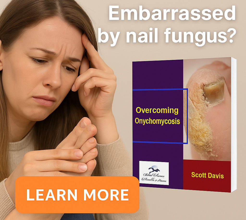
A fungal nail often has a classic look. It may become yellow, white, brown, thickened, brittle, rough, crumbly, or separated from the nail bed. Those clues matter, and they are often what bring a person into the clinic in the first place. CDC, Mayo Clinic, and AAD all describe these kinds of changes as common features of fungal nail infection.
But appearance alone can fool people. Mayo Clinic says psoriasis can mimic a fungal nail infection, and AAD says nail psoriasis or nail injury can sometimes look like nail fungus. AAFP also notes that common mimics include chronic trauma and psoriasis. That is why a nail can look like a fungal invader while actually telling a completely different story.
This is one of the most important truths in the whole topic. A nail is a good storyteller, but not always an honest one. It gives clues, not a verdict.
The first step is the clinical exam 🩺
Diagnosis often begins in the most ordinary way. A dermatologist or other clinician looks carefully at the nails, sometimes both fingernails and toenails, and checks for color change, thickening, buildup beneath the nail, lifting, crumbling, or shape distortion. AAD says dermatologists examine both fingernails and toenails and look for color change and buildup beneath the nail. They also ask about health history and possible exposure, such as whether you may have encountered nail fungus at home, at a gym, or at a pool.
This history matters because fungal nail infection does not live in a vacuum. A person may also have athlete’s foot, sweaty feet, tight shoes, diabetes, poor circulation, nail trauma, or shared wet-surface exposure. Those details do not prove fungus, but they help shape suspicion and guide what kind of sample to collect and how strongly a fungal diagnosis should be pursued. This is an inference, but it is grounded in AAD’s description of history-taking and the recognized risk factors described across CDC and Mayo Clinic sources.
The next step is usually taking a sample ✂️
If onychomycosis is suspected, the clinician often takes material from the nail. AAD says this can include removing buildup from a nail, clipping off a bit of the nail, or scraping the nail surface. Mayo Clinic similarly says a provider may take nail clippings or scrape debris from under the nail, and those samples are sent to a lab.
This is the part many people do not expect. They think diagnosis means somebody glances at the nail and nods wisely. But proper diagnosis usually means actually collecting evidence from the nail itself. In a way, the nail has to testify.
Where the sample is taken matters 🎯
Not every nail sample is equally useful. AAFP’s rapid evidence review says samples should be taken from the most proximal area of onycholysis, meaning the part closest to where the infection is active rather than the most obvious dead edge. It also notes that the nail plate may need to be trimmed back to reveal the right sampling area. Older AAFP guidance adds that, for suspected onychomycosis, the area should be cleaned and then subungual debris and nail clippings should be collected, ideally from the most representative diseased area.
That detail matters because fungus may be living deeper or closer to the active margin, not just in the crumbling tip everyone can see. A poor sample can produce a false-negative result, which means the fungus was there but the test missed it. That is an inference, but it follows directly from the emphasis on good sampling technique in AAFP guidance.
What tests are used? 🧪
Several tests can help diagnose onychomycosis. The most commonly discussed are potassium hydroxide preparation, fungal culture, periodic acid–Schiff staining of nail clippings, and polymerase chain reaction testing. AAFP’s 2021 review says a KOH preparation with confirmatory fungal culture, PAS stain, or PCR is the preferred diagnostic approach. It also states that KOH with direct microscopy is the preferred initial method because it is highly specific, rapid, and cost-effective.
Let’s unpack those one by one, because this is where the laboratory detective work begins.
Potassium hydroxide preparation, or KOH, is the quick classic 🔬
KOH preparation is often the first-line office or laboratory test. In simple terms, nail material is placed with potassium hydroxide so keratin dissolves and fungal elements become easier to see under the microscope. AAFP explains that KOH leaves fungal structures intact and allows identification of hyphae, pseudohyphae, or spores under microscopy. AAFP’s rapid review also says KOH with direct microscopy is generally the preferred initial diagnostic method.
The beauty of KOH is that it is relatively quick and specific. The weakness is that it does not always tell you exactly which organism is involved, and it can miss infection if the sample was poor or the fungal burden is low. That is why negative KOH does not always end the conversation when suspicion remains high. AAFP says if KOH is negative and suspicion remains high, other testing may be done to confirm the diagnosis.
KOH is a bit like shining a flashlight into a cave. You may spot the intruder quickly, but sometimes you need a second tool to be sure who is hiding there.
Fungal culture helps identify the organism 🌱
A fungal culture can be performed using nail clippings or subungual debris. AAFP says culture helps identify the organism and allows species differentiation, though it takes longer and is limited by lower sensitivity and cost. Mayo Clinic likewise notes that samples may be sent to a lab to identify the cause of symptoms.
This means culture can answer a more specific question than KOH: not just “is fungus present?” but “what fungus may be growing?” That can matter if treatment decisions become more complicated or if there is concern about an unusual organism. The trade-off is patience. Culture takes time, often much longer than people expect. AAFP’s older review notes results are usually available in four to six weeks.
PAS stain is often very useful 🧾
Periodic acid–Schiff staining, usually called PAS stain, is done on nail clippings that go to pathology. AAFP says PAS staining is highly sensitive and can help confirm infection. The 2021 review notes biopsy plus PAS stain as one of the more sensitive diagnostic methods, and the 2014 AAFP review says PAS may be the most sensitive diagnostic test, although it is more expensive.
PAS is often especially helpful when you want stronger confirmation and a KOH result is negative or uncertain. It can show fungal elements within the nail tissue itself. AAFP also notes that PAS stain can help assess the degree of nail plate involvement.
In practical terms, PAS is a bit like not only seeing footprints, but seeing them stamped into the floorboards.
PCR is newer and can also confirm the diagnosis 🧬
PCR, or polymerase chain reaction, is another confirmatory option. AAFP’s 2021 review says PCR can confirm the diagnosis but is more expensive than other tests. It offers another way to detect fungal material and can be useful in some settings, though it is not always the first test used in ordinary practice.
So yes, PCR belongs in the diagnostic toolbox, but the classic combination of exam plus KOH and possibly culture or PAS remains more typical in many settings.
Why confirmation matters before treatment 💊
This may be the biggest practical point for patients. Treatment for onychomycosis can be long, sometimes expensive, and sometimes involves oral antifungal medication with meaningful side effects or monitoring needs. AAD’s Choosing Wisely statement says about half of suspected fungal nails are not actually fungal, and it specifically recommends not prescribing oral antifungal therapy without confirmation. CDC also says finger and toenails can change shape or color for many reasons, including previous injuries, and advises talking to a healthcare provider about testing before beginning treatment for a fungal infection.
That means confirmation is not just academic fussiness. It protects people from unnecessary medication and makes it more likely the real problem will be treated correctly.
What conditions can mimic onychomycosis? 🎭
Several conditions can masquerade as nail fungus. Mayo Clinic names psoriasis as one mimic and notes that yeast and bacteria can infect nails too. AAD says nail psoriasis and nail injury can resemble nail fungus. AAFP adds that common mimics include chronic trauma and psoriasis.
So if a nail is thick, yellow, lifted, or rough, the list can include:
psoriasis,
old trauma,
bacterial or yeast infection,
other nail dystrophies,
and true onychomycosis.
That is why a lab-confirmed answer is often worth much more than a confident guess.
Can diagnosis ever be made without testing? 🤷♂️
In some real-world situations, clinicians may sometimes treat based on strong suspicion, especially when testing is not readily available or cost is a major issue. AAFP’s 2021 review says empiric treatment with terbinafine can be considered if testing is cost prohibitive. But the same review and AAD guidance still make clear that confirmatory testing is generally recommended before treatment begins.
So the fair answer is:
sometimes yes, but good practice usually favors confirmation.
A practical diagnosis pathway 🧠
If we turn all of this into a simple real-world pathway, it usually looks like this:
A clinician examines the nail and asks about symptoms, exposures, and health history. If fungus seems likely, they collect nail clippings, subungual debris, or surface scrapings. The sample may then be examined under a microscope with KOH, sent for culture, sent for PAS staining, or in some settings tested by PCR. The final diagnosis is based on the combination of what the nail looks like and what the sample shows.
That is the diagnostic journey in plain language. It is half detective work, half laboratory proof.
So, how is onychomycosis diagnosed? ✅
Here is the cleanest answer.
Onychomycosis is diagnosed by clinical examination plus confirmatory testing of nail material. A dermatologist or clinician examines the nails, asks about health and exposure, and often takes nail clippings, subungual debris, or scrapings. These samples may be tested with KOH microscopy, fungal culture, PAS staining, or PCR. Because many nonfungal nail disorders look similar, confirmation is usually recommended before starting oral antifungal therapy.
So the smartest one-sentence summary is this:
Onychomycosis is diagnosed not by the nail’s appearance alone, but by making the nail hand over evidence.
Final thoughts from the road 🌏
Across Thailand, Laos, Vietnam, Cambodia, Myanmar, India, and many other Asian countries, I have learned that small body problems often hide inside bigger assumptions. A thick yellow nail looks obvious until it is not. A damaged nail looks fungal until the microscope says otherwise. The nail may whisper one story, but the lab sometimes tells a different one.
So if you ask me one final time, how is onychomycosis diagnosed?
My answer is this:
It is diagnosed by looking carefully, sampling the nail, and confirming fungus with testing rather than relying on appearance alone.
FAQs ❓
1. Can a doctor diagnose nail fungus just by looking?
Sometimes a clinician may strongly suspect it by appearance, but confirmation is usually recommended because many nail problems can look similar.
2. What sample is taken to diagnose onychomycosis?
Common samples include nail clippings, debris from under the nail, or surface scrapings from the nail.
3. What is a KOH test for nail fungus?
It is a microscope test in which nail material is prepared with potassium hydroxide so fungal elements are easier to see.
4. Is KOH enough to diagnose onychomycosis?
It is often enough to begin treatment because it is rapid and highly specific, but culture, PAS stain, or PCR may be used when more confirmation is needed.
5. What does fungal culture do?
Fungal culture can help identify the organism causing the infection, but it takes longer and is less sensitive than some other methods.
6. What is PAS stain?
PAS stain is a pathology test on nail clippings that can be highly sensitive for detecting fungal elements in the nail tissue.
7. Why not just start treatment without testing?
Because about half of nails that look fungal may not actually be fungal, and oral antifungal therapy can have side effects.
8. What conditions can mimic nail fungus?
Psoriasis, nail injury or trauma, and some yeast or bacterial infections can mimic onychomycosis.
9. Does the sample location matter?
Yes. AAFP guidance says sampling from the most proximal active diseased area improves diagnostic yield.
10. What is the easiest way to remember the diagnosis process?
Think of it this way: first the nail is examined, then the suspicious material is sampled, then the microscope or lab decides whether fungus is really there.

I’m Mr.Hotsia, sharing 30 years of travel experiences with readers worldwide. This review is based on my personal journey and what I’ve learned along the way. Learn more |